Lacunar Stroke Recovery: Timeline, Evidence-Based Rehabilitation Strategies, and Expert treatment Tips

Quick recognition of lacunar stroke symptoms is critical because timely intervention can dramatically improve outcomes. Stroke remains the second leading cause of death worldwide and is the top cause of long-term disability in the United States. Among all ischemic strokes, lacunar strokes account for approximately 25%, making them a significant public health concern [1].
Although lacunar strokes are small, they can still lead to significant physical, cognitive, and functional challenges that affect daily life and long-term independence. The numbers tell a sobering story – a large portion of patients don't survive the stroke or its complications, while another nearly 20% face another cerebrovascular event [2]. On top of that, a significant portion still struggle with functional impairments even five years later. The good news? People who get emergency treatment have a real shot at full recovery, and they tend to bounce back better than those with other types of strokes.
Recovery after a lacunar stroke is often more favorable than recovery from larger strokes, but outcomes depend on how quickly treatment begins, the location of the stroke, and how consistently rehabilitation is performed. Many people want to know: How long does lacunar stroke recovery take? Can you fully recover? This article answers those questions while outlining evidence-based rehabilitation strategies that support long-term recovery.
This piece dives into what makes a lacunar stroke unique and why its small-vessel location creates specific cognitive and motor challenges that affect recovery. You'll learn about the most effective ways to rehabilitate based on solid evidence, such as early movement and repeated task practice after stroke. We'll also share practical tips backed by therapists to help achieve the best possible outcomes for both lacunar and general stroke recovery.
Understanding Lacunar Stroke and Its Unique Challenges
The term “lacunar” comes from the Latin word lacuna, meaning small cavity or gap. In a lacunar stroke, these small cavities form when a tiny area of brain tissue dies due to lack of blood flow. Although lacunar strokes are small, they occur deep within the brain’s subcortical structures, where critical communication pathways reside.
What is a lacunar stroke?
To understand lacunar strokes, it helps to start with the two main types of strokes:
- Ischemic stroke – caused by a blockage that prevents blood from reaching part of the brain.
- Hemorrhagic stroke – caused by a blood vessel breaking and bleeding into the brain.
Lacunar strokes fall under the ischemic category. However, they are different from the larger and more familiar types of ischemic strokes. A lacunar stroke happens when one of the brain’s small, deep-penetrating arteries becomes blocked. These tiny vessels supply important subcortical structures located beneath the brain’s surface. The areas most commonly affected include:
- Thalamus
- Basal ganglia
- Pons
- Internal capsule and nearby white matter pathways
Because these arteries are small, the resulting infarcts are small as well, generally less than 15 mm in diameter. Even though they affect small areas, lacunar strokes can disrupt major communication pathways responsible for movement, sensation, and coordination. These strokes affect deep brain pathways rather than the cerebral cortex, lacunar stroke symptoms and recovery patterns differ from other ischemic strokes, which has important implications for rehabilitation planning.
How small vessel strokes differ from other types
The small arteries involved in lacunar strokes branch directly from larger, high-pressure vessels rather than gradually tapering. This exposes them to more stress, making them especially vulnerable to damage from high blood pressure.
Most lacunar strokes result from intrinsic small vessel disease, including:
- Lipohyalinosis – degeneration of the vessel wall caused by chronic high blood pressure.
- Microatheroma – tiny plaque buildup that narrows small arteries.
- Fibrinoid necrosis – damage to the vessel wall from severe or long-standing hypertension.
These processes differ from the common causes of cortical strokes, which often involve blood clots traveling from the heart or major arteries. Because of this, lacunar strokes rarely start with clots formed elsewhere in the body.
Why location matters: cognitive and motor effects
The location of a lacunar stroke plays a major role in the symptoms that appear. Because these strokes occur in subcortical regions, they typically do not cause cortical symptoms such as aphasia, neglect, or major visual disturbances. Instead, each lacunar stroke produces a specific clinical syndrome based on which deep neural pathway is affected.
Research shows that lacunar strokes can significantly impact cognitive function. Even when physical symptoms are mild, about half of patients develop subtle cognitive changes. New lacunes commonly affect processing speed, executive function, and fine motor control.
Different small vessel pathways supply different parts of the subcortex, which explains why the exact location of the blockage determines the specific syndrome and why location matters so much for rehabilitation planning.
Diagnosing and Evaluating Lacunar Stroke
Early diagnosis is essential for improving recovery after a lacunar stroke. Because these strokes affect small, deep areas of the brain, recognizing the right symptom patterns and choosing the best imaging tools helps providers act quickly and accurately.
Common lacunar stroke symptoms to watch for
Lacunar stroke symptoms often start suddenly, but in some cases, they may progress gradually over several hours. Unlike cortical strokes, lacunar strokes usually do not cause language problems, vision loss, or neglect. Instead, they produce a set of classic symptom patterns.
The most common lacunar syndromes include:
- Pure motor hemiparesis: sudden weakness on one side of the body involving the face, arm, and leg
- Ataxic hemiparesis: weakness combined with clumsiness or poor coordination
- Pure sensory stroke: numbness or altered sensation on one side without weakness
- Sensorimotor stroke: a combination of weakness and sensory loss
- Dysarthria–clumsy hand syndrome: slurred speech and hand clumsiness
Because these strokes affect deep motor and sensory pathways, these patterns can help clinicians identify a likely lacunar infarct even before imaging. Recognizing these symptom patterns early is critical, as prompt treatment and early rehabilitation significantly improve lacunar stroke prognosis and recovery outcomes.
Imaging techniques: MRI vs CT
Both CT and MRI are used to evaluate stroke, but MRI is much better at detecting lacunar infarcts. CT scans are quick and widely available, but they often miss small, deep strokes, especially in the first hours after symptoms start.
MRI, particularly diffusion-weighted imaging (DWI), is far more sensitive and can detect very small lesions within minutes. Because of this, the American Academy of Neurology recommends diffusion MRI as the preferred test when evaluating a possible lacunar stroke.
The role of neurological exams and lab tests
A careful neurological exam is still one of the most important parts of diagnosing a lacunar stroke. Providers check for facial droop, arm or leg weakness, changes in sensation, speech clarity, and coordination. Simple tools like the F.A.S.T. acronym (Face, Arm, Speech, Time) help quickly identify possible stroke signs.
Basic lab tests are also done early on. These typically include blood glucose, blood counts, electrolytes, and clotting studies. The goal is to rule out other conditions that can mimic stroke, make sure the patient is stable for treatment, and identify any issues that could affect care. An ECG may also be used to screen for heart rhythm problems.
A lacunar stroke diagnosis is made by combining:
- A clinical pattern consistent with a lacunar syndrome
- Imaging that shows no major vessel blockage
- MRI evidence of a small, deep, non-cortical infarct
This combination helps distinguish lacunar strokes from other stroke types and guides the most appropriate treatment and rehabilitation plan.
How Long Does Lacunar Stroke Recovery Take?
Recovery time after a lacunar stroke varies, but many individuals experience meaningful improvements within the first 3 to 6 months, especially with early and consistent rehabilitation. Some people continue to make gains for 12 months or longer, particularly with ongoing task-specific practice and home exercise programs.
Compared to larger strokes, lacunar stroke recovery is often faster and more complete, though subtle motor or cognitive deficits may persist without targeted therapy.
Evidence-Based Rehabilitation Strategies for Recovery
Recovery after a lacunar stroke benefits from targeted, evidence-based rehabilitation that taps into the brain’s ability to reorganize itself, referred to as neuroplasticity. Research highlights several strategies that support motor, sensory, and cognitive improvements.
Early mobilization and its benefits
Getting patients out of bed and moving soon after becoming medically stable is essential. Ideally, beginning rehabilitation 24–48 hours of stroke can lead to significant benefits. Studies show early mobilization helps patients regain independent walking faster. Functional improvements are seen not only at discharge but also at 3- and 12-month follow-ups. Early mobility also reduces complications like pneumonia and blood clots, supporting overall recovery.
Repetitive task practice after stroke
Repetitive task practice involves performing the same meaningful movement or activity over and over to strengthen neural pathways. Repetition is key because it drives experience-dependent neuroplasticity, the brain’s ability to reorganize itself based on repeated experiences. By consistently practicing a specific movement, the brain reinforces the connections controlling that movement, gradually improving strength, coordination, and motor control.
Examples of repetitive task practice commonly used in lacunar stroke rehabilitation include:
- Repeated reaching for and grasping objects to improve hand function
- Step-by-step walking exercises or treadmill practice to enhance lower limb mobility
- Fine motor exercises such as buttoning, stacking coins, or manipulating therapy putty
- Sit-to-stand repetitions to improve leg strength and balance
- Gait training drills focused on step length, foot clearance, and symmetry
Research shows that patients who engage in repetitive task practice not only gain measurable improvements like walking longer distances but also retain these gains for months after therapy. The more meaningful and goal-directed the task, the stronger the neuroplastic response, which directly supports functional recovery.
Constraint-induced movement therapy (CIMT)
CIMT involves restraining the unaffected limb while intensively practicing movements with the affected limb. In some cases, therapists or caregivers will place something on the unaffected to limb to physically limit its use. CIMT focuses on ‘forcing’ the use of the affected limb to further tap into neuroplasticity. This approach:
- Enhances arm and hand function
- Improves daily task performance
- Promotes lasting structural changes in the brain, including increased gray matter in motor areas
Even patients with moderate impairment can benefit when therapy is tailored to their abilities. Constraint-induced movement therapy is often guided by an occupational therapist and is most effective when paired with high-repetition hand and arm exercises, especially during the subacute phase of stroke recovery.
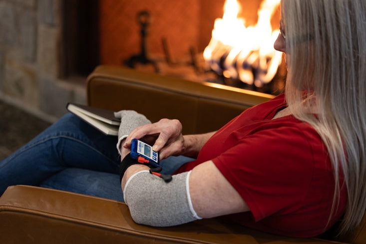
Electrical stimulation for lacunar stroke rehab
Electrical stimulation therapies use small electrical currents to activate muscles or nerves, helping improve movement and support recovery after stroke. These approaches leverage neuroplasticity by repeatedly engaging affected muscles and the brain pathways controlling them.
Types of electrical stimulation commonly used in stroke rehab include:
- FES (Functional Electrical Stimulation): Activates muscles during specific functional tasks, like stimulating ankle dorsiflexors while walking to improve foot clearance.
- NMES (Neuromuscular Electrical Stimulation): Stimulates muscles to produce contractions, usually for strengthening and reducing atrophy, even if the movement is not linked to a specific task.
- SES (Sensory Electrical Stimulation): Targets sensory nerves rather than motor nerves, helping improve sensation and enhance motor learning indirectly.
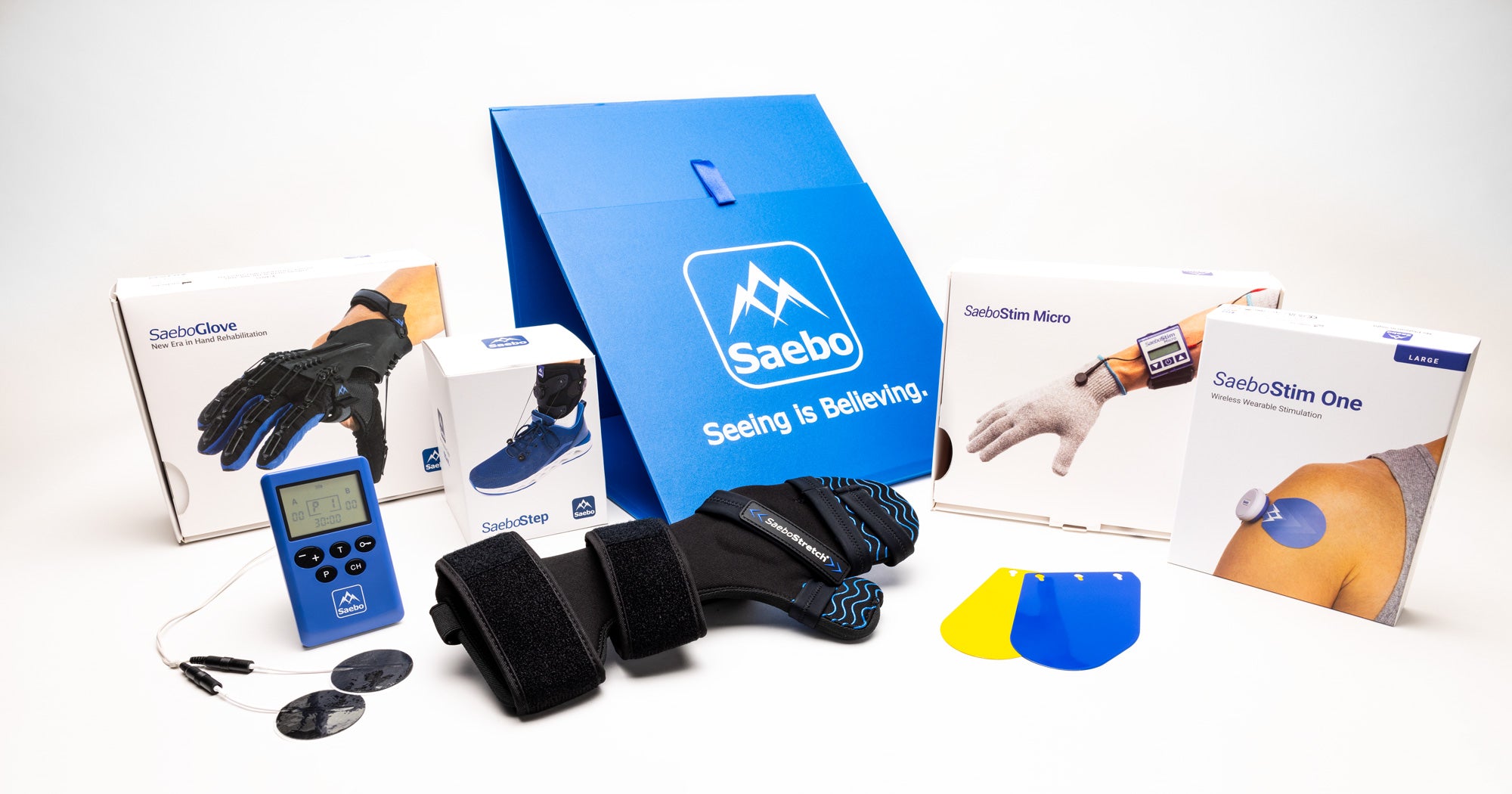
Examples of electrical stimulation devices used in stroke rehabilitation include the SaeboStim Micro, Pro, Spa, and SaeboStim One, which are designed to support neuroplastic, task-specific recovery after stroke. Check out more details here: https://www.saebo.com/
By applying these techniques during repetitive or task-specific practice, therapists can help patients regain strength, coordination, and functional movement. Using a “trigger” function, such as with the SaeboStim Pro, allow you to control the stimulation with voluntary movement, which can be particularly effective for creating more natural, lasting motor patterns.
How to maximize the benefit: Compiling treatment approaches
Recovery after a lacunar stroke is most effective when multiple evidence-based strategies are combined. No single therapy addresses all aspects of motor, sensory, and cognitive recovery, so integrating approaches maximizes neuroplasticity and functional gains.
Practical ways to compile treatments include:
- Early mobilization: Begin movement as soon as it is safe to prevent complications and kickstart recovery.
- Repetitive task practice: Pair meaningful, goal-directed movements with high repetition to strengthen neural pathways.
- Task-specific training: Focus on real-life activities such as reaching, grasping, walking, or balance exercises.
- Electrical stimulation (FES, NMES, SES): Use stimulation to activate weak muscles, reinforce movement patterns, and enhance the effects of task practice.
- Constraint-Induced Movement Therapy (CIMT): Combine with repetitive and task-specific practice for intensive upper limb retraining.
- Technology-assisted tools (VR, AR, Saebo Devices) Integrate immersive and interactive exercises to boost engagement and simulate real-world tasks.
By layering these therapies thoughtfully, therapists can target multiple pathways simultaneously, improving strength, coordination, and motor control while promoting lasting neuroplastic changes. For example, a patient might practice reaching for objects (task-specific training) repeatedly (repetitive practice) while using FES to assist muscle activation. This integrated approach ensures that each therapy reinforces the others, helping patients achieve the greatest possible functional recovery. Combining these approaches creates a comprehensive lacunar stroke rehabilitation plan that supports faster recovery, improved function, and long-term independence.
Expert Tips for Supporting Long-Term Recovery
Recovery from a lacunar stroke goes way beyond the first phase of rehabilitation. Your long-term progress needs a smart game plan and steady support from healthcare providers and family members.
Setting realistic goals with your rehab team
Your recovery milestones need to be specific, measurable, attainable, relevant, and time-bound. When patients take part in goal-setting talks, their rehabilitation results improve. Your care team should work together to create customized recovery goals. This team often includes neurologists, rehabilitation specialists, physical therapists, occupational therapists, and speech therapists. These goals often need tweaking as you make progress.
Incorporating stroke rehabilitation into daily life
Your brain's ability to rewire itself after a lacunar stroke depends on consistency. Make it a habit to do your exercises each day and use real-life examples for treatment. You'll stick to your rehab plan better when you connect it to things you love doing. Keep tabs on your progress to see how far you've come.
Managing fatigue and emotional health
Many stroke survivors deal with post-stroke fatigue, whatever the stroke's severity. This tiredness comes from lifestyle changes, emotional adjustments, and due to many simple tasks needing more energy life can become feeling exhausting. You can handle fatigue better by taking planned rest breaks throughout your day. Watch out for signs of depression too - it often shows up during stroke recovery.
Conclusion
Lacunar strokes may be small, but they present unique rehabilitation challenges that require specialized strategies. Early mobilization ideally within the first 24–48 hours can help patients regain independence more quickly, while repetitive task practice and constraint-induced movement therapy support neuroplasticity and rebuilding of neural pathways. Technology-assisted tools, such as electrical stimulation, can enhance traditional rehabilitation when paired with daily practice and realistic, achievable goals.
Recovery extends beyond the clinic. Incorporating exercises into your daily routine, managing post-stroke fatigue, and supporting emotional well-being all contribute to long-term progress. Heart-healthy habits, including regular exercise and a balanced diet, help reduce the risk of another stroke, which remains a concern even after a lacunar stroke.
While the recovery journey can be challenging, many individuals can fully or nearly fully recover from a lacunar stroke, especially when rehabilitation begins early and remains consistent. Evidence-based strategies such as repetitive task practice, occupational and physical therapy, and technology-assisted rehabilitation—like Saebo’s neuroplasticity-based tools—help support lasting functional improvements. With the right plan, persistence, and support, lacunar stroke recovery can lead to meaningful independence and improved quality of life.
References
All content provided on this blog is for informational purposes only and is not intended to be a substitute for professional medical advice, diagnosis, or treatment. Always seek the advice of your physician or other qualified health providers with any questions you may have regarding a medical condition. If you think you may have a medical emergency, call your doctor or 911 immediately. Reliance on any information provided by the Saebo website is solely at your own risk.
