How Stroke Affects the Brain: Key Regions, Symptoms, and Recovery

The brain controls every aspect of how we move, think, speak, and interact with the world, which is why a stroke or brain injury can affect nearly every area of daily life. When a stroke or traumatic brain injury occurs, the effects depend on which brain regions are damaged, influencing movement, speech, cognition, sensation, and overall stroke recovery outcomes.
This guide provides a high-level overview of the brain’s major areas: the cerebrum, cerebellum, and brainstem; their roles in movement, cognition, speech, and daily function, and how injuries can affect these systems. It also highlights how targeted stroke rehabilitation strategies, guided by brain anatomy and neuroplasticity, can support recovery and help patients regain independence.
Understanding Brain Structure: A Simple Breakdown
The brain is made up of three major parts that work together to control everything we think, feel, and do; and damage to any of these areas can shape the challenges seen in stroke or brain injury recovery. One of the most important areas is the cerebrum, which is responsible for our conscious actions and higher-level thinking. A thick bundle of about 200 million nerve fibers, called the corpus callosum, connects the left and right hemispheres so they can communicate.
The outer surface of the cerebrum is the cerebral cortex which is the wrinkled layer that helps us process information, make decisions, and carry out complex tasks. The cortex is divided into four lobes — frontal, parietal, temporal, and occipital — each responsible for functions commonly affected after a stroke. In addition to the four primary lobes, other regions frequently affected by stroke include the cerebellum and brainstem.
Frontal Lobe
The frontal lobe plays a major role in decision-making, problem-solving, personality, and voluntary movement. It also contains Broca’s area, the primary region responsible for speech production. If this area is affected by stroke, survivors may experience changes in behavior, difficulty speaking, impaired decision-making, or weakness on one side of the body.
Parietal Lobe
The parietal lobe helps us process sensory information, including touch, temperature, and pain. It also supports spatial awareness, helping us understand where our body is in relation to the environment. Injury here may affect sensation, coordination, spatial awareness, or the ability to recognize objects (agnosia).
Temporal Lobe
The temporal lobe is essential for hearing, memory formation, and language comprehension. It houses Wernicke’s area, which helps us understand spoken language. Damage can lead to difficulty understanding words (receptive aphasia), memory problems, or changes in emotional responses.
Occipital Lobe
The occipital lobe is the brain’s visual processing center. It interprets shapes, colors, motion, and visual patterns. It also contributes to facial recognition, working closely with structures in the nearby temporal lobe. Injury can cause visual field loss, visual processing deficits, or difficulty recognizing objects.
Cerebellum
The cerebellum sits at the back of the head underneath the occipital lobe. Even though it makes up only about 10% of the brain’s total volume, it contains more than half of all the brain’s neurons. It plays an important role in coordinating movement rather than starting it.
The cerebellum helps keep movements smooth and accurate by comparing what the body intended to do with the sensory feedback it receives. Injury to this area can lead to ataxia (uncoordinated movement), balance problems, tremors, and difficulty learning new motor skills.
Brainstem and life-sustaining functions
The brainstem connects the cerebrum to the spinal cord and controls many automatic functions that keep us alive. It is divided into three main parts, each with specific responsibilities:
- Midbrain: Helps regulate visual and auditory reflexes
- Pons: Supports breathing, facial movements, and sleep cycles
- Medulla oblongata: Regulates heart rate, blood pressure, and breathing
The brainstem also contains ten of the twelve cranial nerves, which help manage essential functions like swallowing, facial sensation, eye movements, and speech. Damage to this area can seriously affect breathing, heart rate, swallowing, alertness, and other basic survival functions.
How the Cerebrum, Cerebellum, and Brainstem Work Together
The brain’s hemispheres, cerebellum, and brainstem form an interconnected network that allows the body to move, sense, and survive. Broadly speaking, the cerebrum plans and initiates voluntary movements and processes sensory information. The cerebellum refines these movements, ensuring they are smooth, balanced, and coordinated. The brainstem acts as a communication highway, sending signals between the brain and the body while controlling vital automatic functions like breathing, heart rate, and reflexes.
This collaboration explains why stroke symptoms vary significantly depending on the location of brain damage. Damage to one area may affect movement, coordination, or basic life functions, while recovery often depends on retraining the precise circuits that were disrupted.
What Happens When Each Brain Region Is Injured
Brain injuries produce specific patterns of deficits depending on which region is affected. Doctors and their care team use these patterns to identify the damaged areas and guide rehabilitation. The location and severity of the injury determine the types of symptoms a patient may experience.
Stroke on left side of brain: what is damaged?
When a left hemisphere stroke occurs, it primarily affects language processing and the right side of the body. This is because the left hemisphere controls speech, comprehension, and voluntary movement on the opposite side of the body. Common left brain stroke symptoms include:
- Difficulty finding words or understanding speech
- Weakness or paralysis on the right side of the body
- Challenges with calculations or logical thinking
- Signs of depression, cautious behavior, or frustration
The exact symptoms and severity depend on the specific areas of the left brain that are affected. Because the left hemisphere houses the brain’s language centers, strokes here often have a greater impact on daily communication.
Right hemisphere stroke: common symptoms
A right hemisphere stroke creates a different set of challenges. This side of the brain controls spatial awareness and the left side of the body. Common signs of a right-side stroke include:
- Lack of awareness of the left side of the body or environment
- Poor balance and coordination
- Visual changes
- Difficulty recognizing faces or finding objects
- Impulsive behavior and limited insight into their condition
Right hemisphere injuries can make it hard for patients to navigate their surroundings safely, sometimes causing them to bump into objects on the left side.
Injuries to the cerebellum and brainstem
Injuries to the cerebellum or brainstem often produce severe motor and life-sustaining problems.
- Cerebellar damage affects coordination and balance. Patients may develop ataxia, showing wide, staggering steps and tremors, and may have difficulty with precise movements or speech.
- Brainstem damage is particularly serious because this region controls breathing, heart rate, and consciousness. Severe injuries can lead to locked-in syndrome, where a patient is awake and aware but completely paralyzed except for eye movements.
How damage affects thinking, movement, and senses
The effects of a brain injury depend on which regions are involved. Frontal lobe damage can change judgment, behavior, and problem-solving, while parietal injuries affect sensation and spatial awareness. Temporal lobe injuries disrupt memory and language comprehension, and occipital damage leads to vision problems. Injuries to the cerebellum interfere with coordination and balance, and damage to the brainstem can affect vital functions like breathing, heart rate, and alertness.
Movement problems usually appear on the opposite side of the body because most motor pathways cross in the brainstem — a concept known as contralateral control. Knowing how each region contributes to thinking, movement, and sensation helps guide rehabilitation toward the areas most affected.
Connecting Brain Anatomy to Treatment Plans
Understanding how specific brain regions relate to movement, speech, memory, and behavior is essential for effective stroke rehabilitation and brain injury recovery. When medical teams know exactly which parts of the brain are affected after a stroke, traumatic brain injury, or neurological condition, they can design more precise treatment plans that improve functional recovery.
Why knowing the affected area matters
Medical teams must accurately identify brain abnormalities through team assessment to make treatment work. Precise mapping helps identify deficits, guide therapy decisions, monitor progress, and improve functional recovery outcomes. Doctors can develop individual-specific treatment approaches that target damaged neural systems by identifying specific regions affected by stroke or injury.
The brain’s ability to reorganize itself, called neuroplasticity, varies by region and recovery stage. These reorganization abilities are the foundations of rehabilitation strategies. Understanding affected areas helps predict likely deficits and recovery patterns. This knowledge lets therapists prepare for challenges and pick the right interventions.
Choosing the right rehab approach
Rehabilitation after brain injury typically uses three major therapeutic approaches, each chosen based on which brain regions were damaged and the resulting motor, cognitive, behavioral, or speech deficits.
Restorative Therapy: Restoring Lost Function
This approach focuses on rebuilding impaired skills through repetitive, task-specific practice.
Examples include:
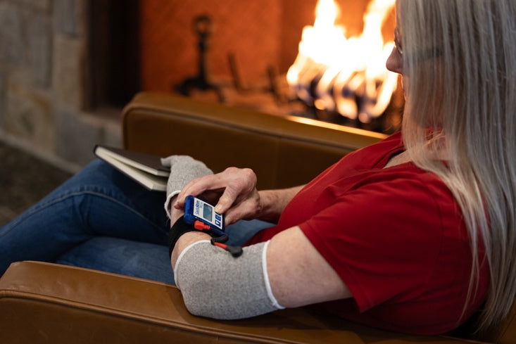
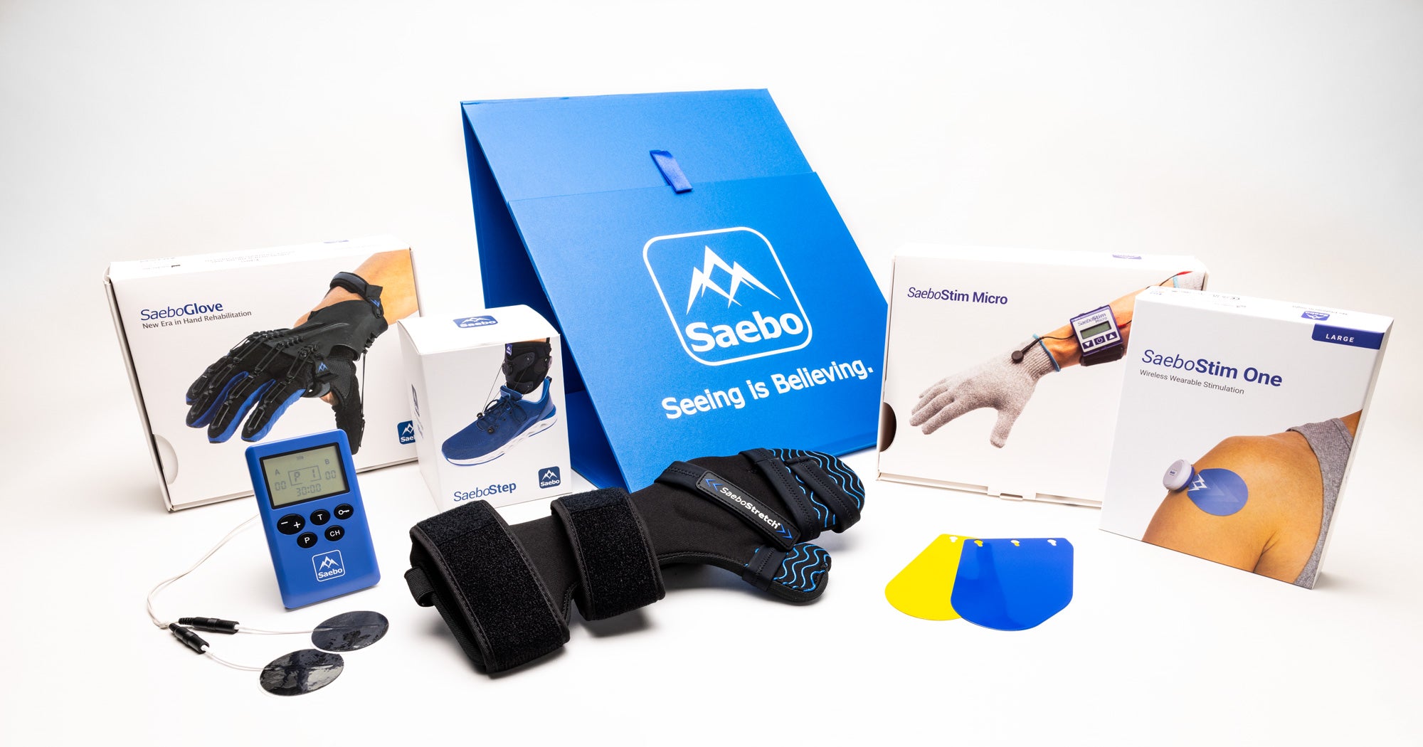
- Repetitive grasp-and-release training using therapy putty, task-specific drills, or assistive devices (such as the Saebo Glove) to improve hand function after stroke.
- Gait retraining on a treadmill with body-weight support to re-establish proper stepping patterns after motor-area damage.
Compensatory Therapy: Working Around Deficits
Compensatory methods help patients function despite ongoing impairments by using tools, strategies, or alternative movements.
Examples include:
- Memory notebooks or smartphone reminders to support individuals with short-term memory loss.
- One-handed dressing techniques for patients who have limited movement or weakness in one arm.
Adaptive Therapy: Building New Routines for Daily Life
Adaptive therapy focuses on helping patients develop sustainable habits and environmental supports that increase independence.
Examples include:
- Home modifications, such as grab bars or adaptive kitchen tools, to enhance safety and accessibility.
- Energy-conservation strategies, such as scheduled rest breaks or simplified routines, for individuals experiencing fatigue or cognitive overload.
The optimal combination of these approaches often evolves as recovery progresses across different levels of care:
- Acute rehabilitation: intensive therapy early after injury
- Post-acute rehabilitation: structured therapy focused on rebuilding strength, coordination, and independence.
- Sub-acute rehabilitation: longer-term, lower-intensity therapy
- Outpatient or day treatment: ongoing support to reinforce skills and build independence
A coordinated care team of physicians, neuropsychologists, speech-language pathologists, occupational and physical therapists, and social workers will work together to deliver comprehensive, patient-centered rehabilitation tailored to everyone’s needs.
Targeted Rehabilitation for Motor, Speech, and Cognitive Recovery
After a brain injury, each affected region requires a customized therapeutic approach. Motor, communication, and cognitive skills recover differently based on the specific neural networks involved, so targeted therapy is essential.
Motor rehabilitation focuses on rebuilding coordination, strength, balance, and motor control after stroke or neurological injury. Injuries to the right or left hemisphere can cause weakness and movement impairments, while cerebellar damage may lead to ataxia requiring specialized interventions. Therapists may use gait retraining, strengthening, task-specific practice, balance training, and adaptive equipment to restore safe mobility.
Speech and language therapy addresses communication challenges such as aphasia and dysarthria. Aphasia, resulting from injury to language centers, requires strategies for improving comprehension, word-finding, and sentence construction. Dysarthria, caused by motor speech impairment, benefits from articulation exercises, breath support training, prosody work, and oral-motor coordination strategies.
Cognitive rehabilitation strengthens information processing, memory, attention, and executive function. Memory training may use restorative techniques (repetition, imagery) or compensatory tools (notebooks, reminders). Executive function deficits often respond well to metacognitive strategy training that builds self-monitoring and problem-solving skills. Compensatory strategies and assistive technologies help patients manage daily tasks more efficiently.
Across all domains, therapy goals are guided by neuroplasticity principles, including repetition, task specificity, intensity, and meaningful engagement.
Conclusion
Understanding brain anatomy is essential for guiding effective stroke rehabilitation and brain injury recovery. Each brain region, the cerebrum, cerebellum, and brainstem, controls specific functions, so the location of damage predicts which abilities are affected, from movement and coordination to speech and cognitive processing. For example, left hemisphere injuries often impact language, while right hemisphere injuries affect spatial awareness, and cerebellar damage disrupts coordination.
This anatomical insight allows medical teams to design targeted rehabilitation plans that improve movement, speech, cognition, and daily function. It also helps patients engage more meaningfully when they understand how exercises relate to the areas of the brain being retrained.
Neuroplasticity — the brain’s ability to reorganize and form new neural connections — offers hope for recovery after stroke. By connecting brain anatomy to symptoms, therapy, and recovery, clinicians and survivors can better understand challenges and pursue more effective rehabilitation strategies.
All content provided on this blog is for informational purposes only and is not intended to be a substitute for professional medical advice, diagnosis, or treatment. Always seek the advice of your physician or other qualified health providers with any questions you may have regarding a medical condition. If you think you may have a medical emergency, call your doctor or 911 immediately. Reliance on any information provided by the Saebo website is solely at your own risk.
