Visual Field Cut vs Neglect After Stroke: Diagnosis, Rehab, and Functional Impact

Stroke survivors may experience visual field cuts (hemianopia) or spatial neglect and correctly distinguishing between them is critical for effective cerebrovascular accident (CVA) rehabilitation and daily function. Visual field cuts affect 20–50% of stroke patients, while spatial neglect impacts a third of right-hemisphere strokes and about 10% of left-sided strokes [1].
While they may appear similar, the two conditions differ fundamentally: visual field cuts cause actual vision loss, whereas hemineglect reflects reduced awareness of one side of space. Accurate identification is essential, as only about 40% of people experience spontaneous recovery within the first 2–3 months after CVA. This guide highlights the differences, their impact on daily life, and evidence-based assessment and rehabilitation strategies to support better clinical decisions.
Understanding the Core Differences in Brain Lesions
Differences in brain structure and neural pathways explain why strokes produce distinct effects such as visual field cuts versus spatial neglect. These two conditions come from damage to specific neural pathways and brain regions. This creates unique challenges that both patients and clinicians must face.
Right hemisphere stroke and left-sided neglect (hemineglect)
Damage to the right hemisphere often causes left-sided hemineglect due to disruption of the brain’s ventral attention network (VAN). The ventral attention network (VAN) in the right hemisphere plays a vital role in spatial awareness, which is your ability to judge distance and depth such as when reaching or navigating a room. Damage to this area makes people develop a strong bias toward the right side of space since they become less aware of the left side. This explains why a third of right hemisphere stroke survivors develop left-sided attention deficits compared to just 10% with left hemisphere damage.
Left-sided neglect most often occurs after damage to key attention areas in the right hemisphere, especially the posterior parietal cortex and the temporoparietal junction. Other affected areas include the superior temporal gyrus, middle frontal gyrus, insula, and basal ganglia. Common symptoms include:
- Their eyes tend to look right without realizing it
- They may miss information or objects that are to their left side
- They struggle to care for their body's left side
- They miss words on the left while reading
Unlike visual field loss, neglect is primarily an attention deficit, not a vision problem, and many patients are unaware of their missing awareness. They don't actively ignore the left side - their brain simply doesn't register it exists.
Posterior visual pathway damage and hemianopia
Homonymous hemianopia differs from spatial inattention in that it reflects actual vision loss, not attentional neglect. It occurs when the visual pathways behind the optic chiasm are damaged, such as the optic tract, optic radiations, or occipital lobe. This causes loss of the same half of the visual field in both eyes. For example, a CVA in the left hemisphere leads to right-sided visual field loss in both eyes (right eye’s temporal field and left eye’s nasal field). Unlike one-sided inattention, hemianopia is a true visual deficit, and most individuals are aware of their vision loss.

Visual field defects show up in 20-57% of post-stroke patients [2]. The location of the damage determines how common these defects are. Here are the most common areas that cause hemianopia:
- Occipital lobe
- Optic radiations
- Lateral geniculate nucleus
- Optic tract
Different brain regions produce distinct visual deficits: temporal lobe injuries often affect the upper visual field, while parietal injuries tend to affect the lower visual field. Unlike neglect, those with hemianopia are typically aware of their vision loss and can learn compensatory scanning strategies.
Overlap and co-occurrence in stroke patients
Visual field loss and spatial neglect frequently co-occur after neurological injury, complicating both diagnosis and rehabilitation planning. Approximately one-third of stroke survivors experience vision loss, with many also showing symptoms of spatial neglect.
Over time, key differences emerge. People with pure hemianopia typically scan toward their blind side in an organized way, adjust their head and eye position to maximize vision, are aware of the problem, and actively try to compensate. In contrast, individuals with hemineglect mainly explore the healthy side of space and fail to attend to the neglected side, even though their primary vision pathways are intact.
Both conditions impact daily life differently: visual field cuts reduce vision in specific regions, whereas lack of awareness disrupts attention to an entire side of space.
Functional Impact on Daily Activities
Stroke survivors experience completely different functional challenges based on whether they have a visual field cut or spatial neglect. These conditions affect daily activities differently and create unique barriers to independence and safety.
Visual field cut: reading, mobility, object finding
People with visual field loss struggle with activities that require full visual scanning. Reading becomes difficult because they can no longer take in a wide sweep of text at once and must learn to make deliberate saccades toward the side of the field loss. They may miss letters, words, or entire portions of text, leading to slower reading and reduced comprehension.
Mobility can also be affected. Individuals may bump into objects, misjudge steps or curbs, and feel unsafe in busy environments. Many adopt compensatory head turns to bring more of their surroundings into view, which can increase neck strain over time.
Finding objects becomes more challenging as anything located in the blind field cannot be seen without turning the eyes or head. Everyday tasks like locating items in the kitchen or navigating crowded spaces require structured scanning strategies that must be learned and practiced.
Spatial neglect: dressing, grooming, navigation
Spatial neglect creates major self-care challenges through what looks like selective inattention. Unlike vision loss on one side, these individuals act as if one side of space no longer exists. This guides them to asymmetric personal care where they might shave, wash, or apply makeup to only one side of their face. During meals, they eat food only from one side of the plate and often complain about getting half-sized portions.
Getting dressed becomes problematic because patients might ignore clothing on the affected side. They try to put both legs into one pant leg or leave one arm without clothes. The same happens with grooming, as they comb, brush, or clean only the non-neglected side.
Moving through space brings more challenges as people drift toward their non-neglected side. In wheelchairs, they have trouble moving toward the inattentive side and often hit objects, doorframes, or walls on that side. Even in bed, they usually position themselves looking away from their neglected side.
Anosognosia and safety risks in neglect
Many patients with spatial awareness deficits also develop anosognosia, meaning they are unaware of their own deficits. Studies show that around 30–60% of CVA survivors with neglect fail to recognize limitations in dressing, navigating, and managing personal belongings [3]. This lack of awareness creates serious safety risks, including:
- Ignoring life-threatening conditions (e.g., chest pain) on the neglected side
- Attempting dangerous activities without assistance
- Increased risk of falls and collisions
- Crossing streets without checking for traffic on the neglected side
- Burns from hot objects on the neglected side
When spatial neglect and anosognosia occur together, the individual cannot use compensatory strategies because they don’t recognize their limitations. This increases caregiver stress and burden.
Both conditions reduce quality of life, but they require different rehabilitation approaches:
- Field cuts → compensatory strategies
- Neglect → attentional training
Accurate identification of each condition is critical to developing effective safety and rehabilitation strategies.
How to determine if its neglect or field cut
Doctors need to distinguish visual field loss from spatial neglect to plan rehabilitation properly. Both cause patients to miss information on one side, but the reasons are very different, and the treatments differ.
Basic screening methods for outpatient therapy can help guide the doctor:
- Visual search patterns: Patients with left-sided neglect start scanning from the right and rarely reach the left side. Those with visual field cuts scan systematically across both sides.
- Line bisection test: Neglect patients mark the center far to the right, while those with left hemianopia mark it slightly left.
- Drawing or copying tasks: Neglect patients omit details on the affected side when drawing with eyes open but improve with eyes closed. Field cut patients do not show this change.
- Observation of daily tasks: Neglect patients may shave or dress only one side or skip words while reading, unlike patients with field deficits who notice and compensate.
Key distinguishing feature: Neglect patients often don’t realize they have a problem (anosognosia), whereas Hemineglect with field cuts are aware of their limitations.
Because about a significant portion of stroke patients may have both conditions, the final diagnosis and treatment plan must be made by the patient’s doctor, often using detailed neuropsychological testing. Correct identification ensures the right therapy—attentional training for neglect or compensatory strategies for visual field loss.
Rehabilitation Strategies Based on Condition Type
The right treatment for stroke survivors depends on their specific visual problems. Visual field loss and neglect need different approaches because they affect the brain in unique ways.
Visual field cut: scanning, reading, and compensatory aids
Those with hemianopia need strategies to compensate for missing visual information. These deficits are not going to ‘go away’ which requires adaptive methods and compensatory techniques in daily life. Some treatment ideas include:
- Anchoring techniques: Use bright markers, colored tape, or contrasting objects at the edge of the visual field to guide scanning.
- “I Spy” or visual search games: Encourage patients to locate targets in their blind field in a structured, engaging way.
- Scanning before head turn: Train patients to move their eyes systematically into the affected field before turning the head.
- Tracking and movement exercises: Follow moving targets with eyes to improve smooth pursuit and visual attention.
These exercises improve reading, navigation, and object detection, helping patients safely explore their environment.
Neglect: prism adaptation, OKS, limb activation
Patients with neglect often ignore one side of space, so therapy focuses on increasing awareness and stimulating the affected side. The goal is ‘retraining’ the individual to include their neglected side back into their daily life. Some example exercises include:
- Placing items on the neglected side: Encourage individuals to reach, grasp, or interact with objects on the affected side.
- Using mirrors: Reflect neglected space to provide visual feedback and increase attention.
- Positioning the therapist or cues on the neglected side: Helps people orient toward the affected side during therapy.
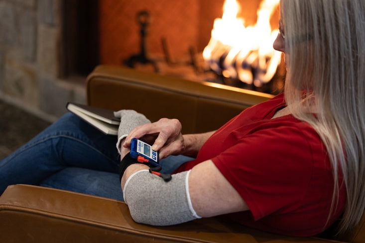
- Increasing stimulation to the affected side: Apply weight, touch, vibration, or use sensory devices like SaeboStim Micro to increase input to the neglected side.
These strategies retrain the brain to attend to and interact with neglected space, improving daily function and safety.
Guidelines for Clinical Decision-Making
Effective management of visual deficits after stroke requires a solid understanding of how visual field deficits and neglect present, so healthcare providers can make appropriate referrals and select targeted interventions.
Ready for discharge- Who to refer to?
Stroke survivors with visual problems may need referrals to several specialists depending on their symptoms. Vision specialists (optometrists or ophthalmologists) are important for people with blurry vision, flashes, black spots, or dimming visual fields, and all stroke individuals should have a vision screening before discharge. Neuropsychologists help identify cognitive or attentional deficits, distinguish them from depression or dementia, and assess decision-making ability and safety risks, such as driving. Occupational therapists can provide functional assessments and therapy recommendations to address daily living challenges caused by neglect or field cuts.
Therapy selection depends on neglect subtype
The specific neglect subtype guides rehabilitation planning. Reading tasks improve with visual scanning training, but it barely helps with non-visual neglect behavior. Each type of neglect needs its own approach - personal, peripersonal, and extrapersonal neglect all require different treatments. Recovery rates change by subtype too. Personal and extrapersonal neglect patients show better recovery after six months compared to those with peripersonal neglect.
Treatment plans need regular updates
Treatment monitoring should focus on functional outcomes rather than impairment assessments. Standard tools help track progress and determine if changes are needed. Driving safety checks must happen regularly for those with visual field defects, following licensing rules. Doctors can adjust treatment intensity or try combining different approaches if progress slows down.
Conclusion
Healthcare professionals must distinguish between visual field cuts and spatial neglect to provide effective rehabilitation for stroke survivors. Field cuts refer to actual loss of vision in part of the visual field, while neglect makes patients unaware of one side of space despite intact vision, often leading to anosognosia. These conditions impact daily life differently: field loss may affect reading, navigation, and object finding, whereas neglect disrupts dressing, grooming, and mobility.
Accurate assessment using visual search patterns, line bisection, and observation across situations is essential, especially since about 30% of patients may have overlapping conditions. Treatment should match the problem: field cuts benefit from scanning training, reading strategies, and compensatory tools, while neglect may need more awareness and body positioning practice. Referrals to the right specialists—vision experts for field cuts and neuropsychologists for complex neglect—help tailor therapy. Recognizing the differences allows targeted interventions, improves functional outcomes, and supports greater independence for stroke survivors.
References
All content provided on this blog is for informational purposes only and is not intended to be a substitute for professional medical advice, diagnosis, or treatment. Always seek the advice of your physician or other qualified health providers with any questions you may have regarding a medical condition. If you think you may have a medical emergency, call your doctor or 911 immediately. Reliance on any information provided by the Saebo website is solely at your own risk.
